Dermoscopy Explained: How Doctors See Beneath the Skin
Dermoscopy, sometimes called dermatoscopy, is a simple, painless way for doctors to take a closer look at the skin. Using a small handheld device called a dermatoscope, your doctor can see details beneath the surface that aren’t visible to the naked eye. This extra level of detail makes dermoscopy one of the most important tools in detecting skin cancer early and understanding other skin conditions more accurately.
How Dermoscopy Works
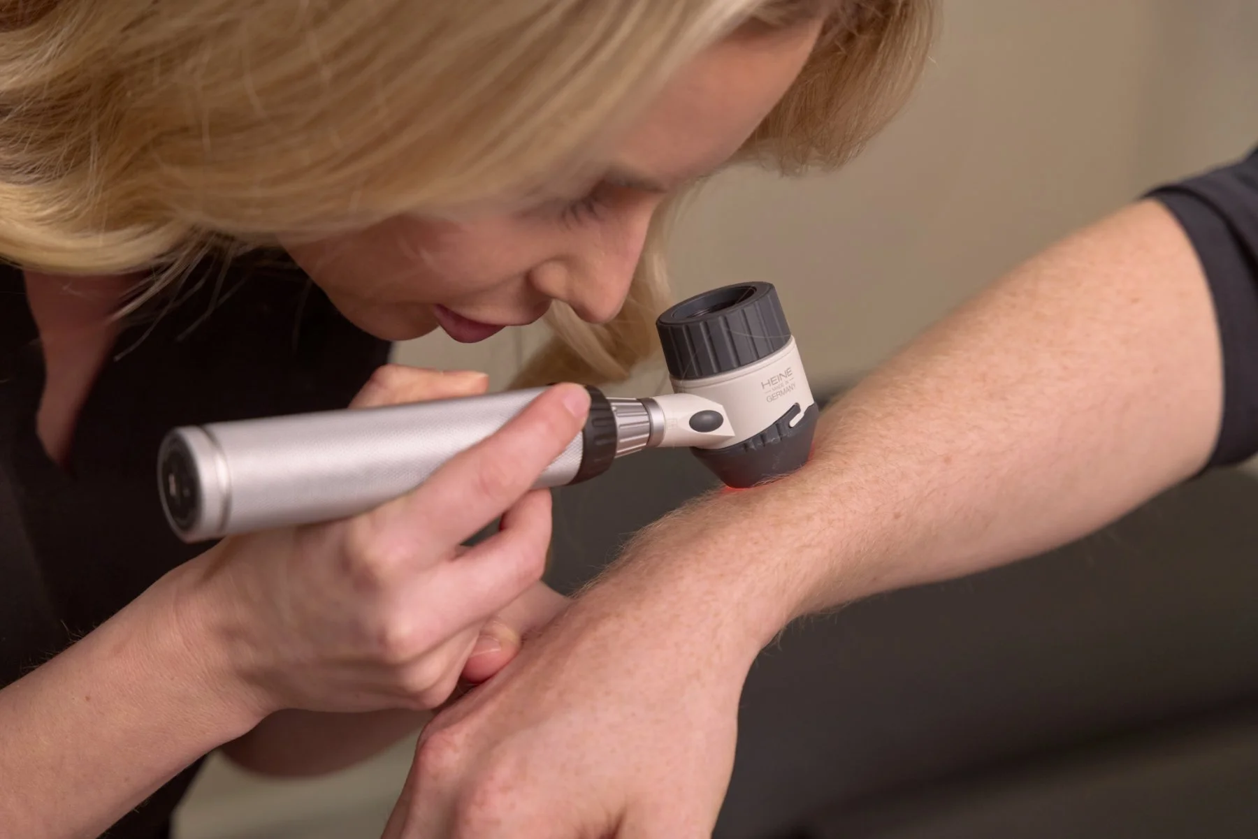
A dermatoscope is like a specialised magnifying glass with built-in light. When placed against the skin, it removes glare and highlights structures just below the surface. Some dermatoscopes use a gel or oil to reduce reflection, while others use polarised light to achieve the same effect.
During a dermoscopic check, your doctor looks at things such as:
The edges or borders
Variations in colour
Pigment networks and other patterns
Small structures like dots, globules or vessels
These features help distinguish harmless spots (such as common moles or seborrhoeic keratoses) from potentially dangerous ones like melanoma or basal cell carcinoma.
Image: Dr Emily Alfonsi using her dermatoscope during skin check
Why Dermoscopy Matters
For patients, dermoscopy offers peace of mind. It allows doctors to detect suspicious changes earlier and with greater accuracy. Research shows that using a dermatoscope improves the chances of picking up melanoma and reduces the need for unnecessary biopsies.
Some of the key benefits include:
Early detection of skin cancer – spotting changes sooner means treatment can start earlier, improving outcomes.
Fewer unnecessary procedures – clearer detail helps avoid biopsies when a lesion is clearly benign.
Ongoing monitoring – images can be stored and compared over time, making it easier to track any changes if you have many moles or are at higher risk.
Beyond Skin Cancer
While dermoscopy is best known for detecting melanoma and other skin cancers, it is also useful in other areas of dermatology. Doctors may use it to:
Assess inflammatory conditions such as psoriasis or lichen planus
Examine the hair and scalp (known as trichoscopy)
Investigate nail changes (onychoscopy)
Help diagnose certain infections such as scabies or fungal skin disease
What to Expect in a Dermoscopy Exam
A dermoscopy check is quick, painless and part of a standard skin examination. Your doctor will place the dermatoscope gently against your skin and study the spot in detail. If needed, photos can be taken to compare over time.
This is especially valuable for people with a personal or family history of skin cancer, a high number of moles, or lesions that look unusual.
With advances in digital dermoscopy, doctors can now capture high-resolution images and even use computer-assisted tools to support diagnosis and monitoring.
Conclusion
Dermoscopy has transformed the way doctors assess the skin. By providing a detailed view beneath the surface, it helps detect skin cancer earlier, improves accuracy and reduces unnecessary procedures. If you’ve noticed a new spot, a change in an existing mole, or if you’re at higher risk of skin cancer, book a skin check with your doctor and ask about dermoscopy.
Written by Dr Emily Alfonsi
MBBS, FRACGP, DRANZCOG
Medical Director, Shade Skin
Dr Emily is a skin cancer doctor with advanced training in diagnosis and treatment. She has personally detected and treated hundreds of skin cancers and is passionate about early intervention and patient education.

